We and our partners use cookies and other tracking technologies to collect data relating to you to perform analytics, improve your experience of using our website, provide you with personalized ads and content based on your interactions with these and other websites and allow you to share content on social media. By clicking “Accept All Cookies”, you consent to this and to the sharing of this data with our partners. You can change your consent preferences at any time in the “Cookie Settings” section at the bottom of our website. Review our Cookie Notice to learn more about our practices.
细胞外囊泡(EV)
按需灵活定制您的细胞外囊泡工作流程
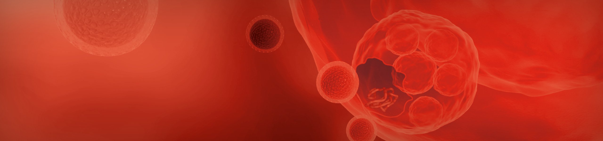
近期,细胞外囊泡(EV)因作为治疗靶点和生物标志物,在疾病的诊断与治疗方面具备巨大的研究潜力而备受关注。这些囊泡是由几乎所有细胞类型释放,并通过转运 mRNA 和 miRNA 在细胞间通讯中发挥关键作用。
细胞外囊泡的多样性特征,为研究人员开辟了众多相关领域的研究和开发机遇。
然而在保持其囊泡和内含物完好无损的同时,可靠地分离出细胞外囊泡仍是一项重大挑战。由于缺乏标准化的分离和纯化方法,加之细胞外囊泡固有的异质性,使得生产和纯化过程更为复杂。
依赖通用解决方案来开发针对您特定需求的EVs分离和分析工作流程是相当困难的。我们独创的工作流程可为您灵活定制实验方案,助您加速获取所需答案,进一步挖掘所需细胞外囊泡种类的研究潜力。
 |
|
 |
|
 |
|
 |
   |
 |
 |
   |
|||
|
细胞培养 |
自细胞、培养基中分离 |
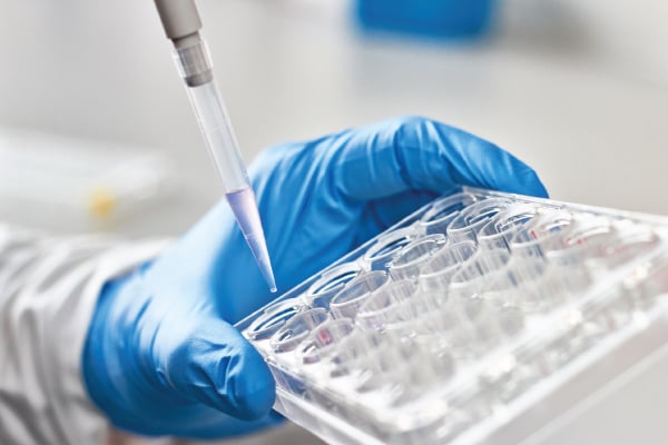
纯化 |
囊泡分析 |
|||
| 自动化细胞培养、活率监测和培养基分析解决方案助您加速获得生产用细胞外囊泡。 | 高效制备培养上清液,并从细胞碎片和大颗粒中分离所需微粒。 | 从异质样品中灵活纯化所需大小和密度的细胞外囊泡。按需灵活定制的实验方案,全方位满足您的特定需求。 | 挖掘经纯化的细胞外囊泡研究潜力:分析小颗粒以研究物理特性,提取 RNA 以了解囊泡内含物。 |


什么是细胞外囊泡?细胞外囊泡的作用是什么?

EV 的粒径范围通常在 30 至 500 nm, 可携带多种内含物,并在细胞间通讯和生理过程中发挥关键作用, 包括血管生成、细胞分化和肿瘤生长。 下载海报以了解更多信息。
下载海报


了解有关细胞外囊泡表征的更多信息
|
使用单囊泡流式细胞术 (vFCTM) 和CytoFLEX在复杂生物流体中直接测量 EV —— John Nolan 博士 Cellarcus Biosciences首席执行官
|
 |

是否兴趣了解如何加速推进您的细胞外囊泡研究进程?
如有否有疑问或对演示感兴趣,我们将随时您提供帮助。
