Chargaff:
在建立从细胞中分离凝血因子的离心方案时,发现“高速离心沉淀物的加入,可大幅缩短血浆上清液的凝血时间”。

1945-1979
1. 揭开凝血奥秘:EV 研究早期阶段
在建立从细胞中分离凝血因子的离心方案时,发现“高速离心沉淀物的加入,可大幅缩短血浆上清液的凝血时间”。
在研究中发现了一种颗粒物组分,该组分“经 31,000 xg 高速离心后沉降,且具有显著凝血功能”。他们认为,该组分除了含有促凝血因子外,还含有“多种微小的血细胞分解产物”。
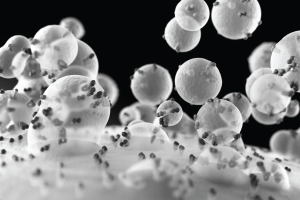
发现了血小板微粒,并将其描述为“一种微小的颗粒状物质,可通过高速离心沉降,虽源自血小板,但与完整血小板不同”。其研究使得人们首次获得了 EV 的电子显微镜图像。
其研究表明EV含有脂质,并携带ATP和收缩蛋白等货物。
发现冬眠唤醒期间蝙蝠甲状腺中存在小的细胞外囊泡。同时在顶膜附近观察到多囊泡体(MVBs),并提出MVB的外膜与顶质膜的融合可能导致囊泡释放到管腔空间。
发现从细胞中释放的囊泡结构并非哺乳动物所独有。例如,褐藻,一种有鞭毛的藻类中,他们同样观察到囊泡从细胞中出芽产生,并通过离心分离出这些EV。同时,EV 还可由热带念珠菌、棒状杆菌、不动杆菌、大肠杆菌和其他物种所释放。
1956-1998
2. 揭开EV的谜团:积累证据,作用不明
在此期间,研究人员积极探寻可能引发疾病(如感染和癌症)的“病毒样颗粒”。然而,研究指出,将从上皮细胞相关的多囊泡体和微囊泡中天然形成的囊泡形态的结构冠以“病毒样”的称号是缺乏合理依据的。
以网织红细胞的成熟过程为模型开展研究,证明腔内囊泡是从细胞中释放而出,并将这些囊泡定义为“外泌体”,即多囊泡体与质膜融合后,经多囊泡体腔内向外释放的颗粒。这一发现揭示了外泌体的分泌途径。
其研究揭示,在细胞外囊泡(EV)与红细胞(RBC)中,脂质和蛋白质在囊泡膜中的横向扩散存在差异。相比红细胞膜, EV 中的蛋白质含量更低,且脂质组分具有随机性,可能导致在 EV 中扩散能力更强。
其研究表明,从网织红细胞中释放出的外泌体保留了酶活性。
其研究揭示牙龈卟啉单胞菌(Porphyromonas gingivalis)细胞外囊泡的产生,以及它们与人多形核白细胞相互作用。
根据不同时期 EV 组分的内吞和脱落情况,Johnstone认为外泌体是废弃膜蛋白分泌的主要途径。该发现对 EV 的释放仅仅是废物清理机制的看法提出了质疑。
其研究帮助人们更好地理解囊泡转运,包括 EV 组分。
其研究揭示,来自免疫细胞的EV 能够呈递抗原。这一研究结果使人们对 EV 的作用有了更深入的理解,尤其是对基于 EV 的治疗策略开发具有重要意义,比如癌症治疗(如Zitvogel 于 1998 年证实)。
其研究证实 EV 组分中存在四次跨膜蛋白(Tetraspanins)。
3. 破解 EV 功能奥密: 揭示 EV 在细胞中的作用
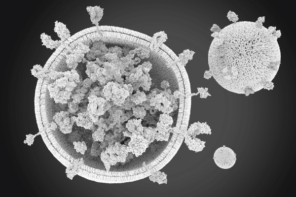
随着人们对细胞外囊泡(EV)在生理过程和疾病发生发展中的作用机制及应用前景的广泛研究,二十一世纪初,该领域研究迎来重大转折点。科学家们开始从蛋白质组学、脂质组学、基因组学、生物化学等多个角度研究 EV 的内含物。这些研究揭示了 EV 在不同情况下的潜在作用。 Chaput(2003)和 Zitvogel(2005)以免疫细胞为对象的研究结果,令癌症免疫疗法领域的研究人员深受启发。Ratajczak(2006)证明细胞源性微囊泡可通过 mRNA和蛋白质水平的转运对其他细胞进行重新编程。这一突破性发现突显了 EV 可能对细胞行为产生的重大影响。 当前,已有证据表明,EV 在免疫应答调节方面具有多种重要作用。(图 1.另请参阅 Buzas,Nature Reviews Immunology,第 23 卷,第 236-250 页(2023 年)。

图 1: EV 的调控功能。位于中央的 EV 在其表面表达特异性标志物, 并与 T 细胞、自然杀伤细胞(NK)和树突细胞(DC)等各种免疫细胞相互作用。调节性 T 细胞(Treg)、细胞毒性 T 淋巴细胞(CTL)、 三磷酸腺苷(ATP)和单磷酸腺苷(AMP)均参与这些相互作用。 请注意,为方便观察,图中所示细胞外囊泡被放大处理。
4. 填补EV 研究领域空白
EV或非EV
目前,科学界中有部分科学家使用“外泌体”一词,而最初这些结构被称为“微粒”。这种命名上的差异容易令人混淆,特别是基于粒径特征任意划分囊泡类型的做法,令这一问题更为突出。为解决这一乱象,某些人士建议采用“细胞外囊泡(EV)一词作为各种由脂质双分子层构成、不能自我复制颗粒的总称。2011年,国际细胞外囊泡学会(ISEV)成立,极大推动了业界对这一术语达成全面共识。在《细胞外囊泡研究基础信息指南(MISEV)》(2018)中,正式确定细胞外囊泡(EV)的定义,即由脂质双分子层构成、不能自我复制的颗粒。学会官方期刊《细胞外囊泡杂志》进一步推动了业界对这一术语的认可,为该领域研究提供了交换数据、想法和信息的平台。
彩页下载
二十一世纪初,该领域研究迎来重大转折点。科学家们开始从蛋白质组学、脂质组学、基因组学、生物化学等多个角度研究 EV 的内含物。这些研究揭示了 EV 在不同情况下的潜在作用。下载彩页了解更多信息
彩页下载
确保可重现性
选择哪种细胞外囊泡(EV)分离和富集方法一直是 EV 领域争论的主要话题。可用技术有多种,包括超速离心法(ultracentrifugation,UC)、超滤法(ultrafiltration)、尺寸排阻色谱(size exclusion chromatography,SEC)、沉淀法(polymer precipitation)、免疫亲和层析(immunoaffinity chromatography)、免疫磁珠捕获(magneto-immunocapture)、微流控免疫芯片(microfluidic immunochips,)和脂质纳米探针(lipid nanoprobes)。选择何种方法主要取决于具体研究目标。
在 EV 表征方面,已开发出多种生物物理方法,如纳米颗粒跟踪分析和脉冲传感技术,均可用于 EV 计数和粒径测定。进一步表征涉及蛋白质组学、基因组学、脂质组学等方法。这些方法均可提供整个 EV 群体的表征信息,而流式细胞术支持对单个 EV 进行表征。单个EV 颗粒的表征信息在揭示不同 EV 的异质性方面具由极高价值。然而,确保研究结果的准确性和可重复性同样至关重要。因此,学术界各位同仁于 2011 年共聚一堂,成立国际细胞外囊泡学会 (www.isev.org), 对推进全球 EV 研究发挥了关键作用。
2018年,该学会发布 EV 分析最新指南《细胞外囊泡研究基础信息指南》(MISEV),旨在解决业内 EV 研究面临的争议和难题。Ramirez 等人表示,“持续关注流式细胞术的原因是它能够可靠区分携带和未携带特定蛋白标志物的 EV,从而准确测定特定类型 EV 的占比”。对样本内 EV 异质性的定量和定性分析,是获取全面信息的关键所在。为了优化这一过程,近期我们还提供了有关使用流式细胞术进行 EV 研究的新资源(Welsh JA、Arkesteijn GJA、Bremer M 等人。单细胞外囊泡流式细胞术汇编。J Extracell Vesicles. 2023;12(2):e12299. doi:10.1002/jev2.12299)。
5. 结论
细胞外囊泡(EV)研究已发展成为一个独立研究领域,已成立专门学会,且定期召开学术会议。EV 的发现向科学界打开了一扇通往未知领域的大门,它在各种生理过程调控中发挥至关重要的作用。进一步开展该领域的研究,有望揭示EV的 异质性,阐明其各种功能,还可能有助于发现新的疾病标志物。通过监测细胞特异性 EV,研究人员可寻找 EV 表面或内部特定标志物。这种方法仅需一次抽血即可实现疾病的监控,已在阿尔茨海默症检测中得到验证。(Tao-Ran (2019)。
此外, EV 可以作为递送载体将治疗用化合物输送至靶细胞或器官的特性,目前正受到研究人员的广泛追捧。研究人员希望通过改造出可靶向特定细胞(比如肿瘤)的载药 EV,攻克传统疗法存在的局限性。这种有针对性的药物递送方法或可提高疗效,尽可能减少不必要的副作用。这一点至为重要,因为目前的疗法通常采用全身给药的方式,不仅疗效欠佳,还可能损害人体健康组织。
参考文献
您是否有兴趣了解如何加速 EV 研究?
如您存在任何疑问或对我们的产品感兴趣,欢迎随时与我们联系,我们将竭诚为您提供帮助。
© 2000-2026 贝克曼库尔特国际贸易(上海)有限公司保留最终解释权

NOT ALL PRODUCTS ARE AVAILABLE IN ALL COUNTRIES.
PRODUCT AVAILABILITY AND REGULATORY STATUS DEPENDS ON COUNTRY REGISTRATION PER APPLICABLE REGULATIONS
The listed regulatory status for products correspond to one of the below:
IVD: In Vitro Diagnostic Products. These products are labeled "For In Vitro Diagnostic Use."
ASR: Analyte Specific Reagents. These reagents are labeled "Analyte Specific Reagents. Analytical and performance characteristics are not established."
CE: Products intended for in vitro diagnostic use and conforming to European Directive (98/79/EC). (Note: Devices may be CE marked to other directives than (98/79/EC)
RUO: Research Use Only. These products are labeled "For Research Use Only. Not for use in diagnostic procedures."
LUO: Laboratory Use Only. These products are labeled "For Laboratory Use Only."
No Regulatory Status: Non-Medical Device or non-regulated articles. Not for use in diagnostic or therapeutic procedures.
![]() 沪ICP备18036651号-1
沪ICP备18036651号-1
![]() 沪公网安备 31010502004935号
沪公网安备 31010502004935号
互联网药品信息服务资格证书-沪网药信备字[2025]00254号
沪(浦)应急管危经许[2022]200234