CD1a Antibodies
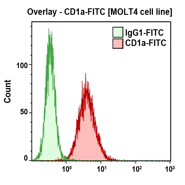
CD1a is a member of the lipid antigen presenting molecules family. Like the other CD1 proteins (group 1: CD1a, CD1b, CD1c and group 2: CD1d, CD1e), CD1a is a transmembrane protein of 49 kDa, noncovalently associated with β2-microglobulin. As other group 1 CD1 antigens, CD1a is found on professional antigen-presenting cells (APC) such as dendritic cells, particularly on Langerhans cells and their direct circulating precursors. It is expressed by those cells related to dermal dendritic cells, derived from monocytes cultured for 6 days in the presence of GMCSF and IL-4. The CD1a antigen is also present on a B cell subset. It is also strongly expressed by cortical thymocytes. CD1a antibodies are useful in the definition of the thymic stage of T cells.
| Clone: SFCI19Thy1A8 (T6) | Isotype: IgG1 Mouse |
| T6 (ref 6T-CD1.7) was used as a CD1a reference monoclonal antibody during HLDA 6. | |
| Clone: BL6 | Isotype: IgG1 Mouse |
| The BL6 antibody recognizes the C epitope of the CD1a molecule. | |