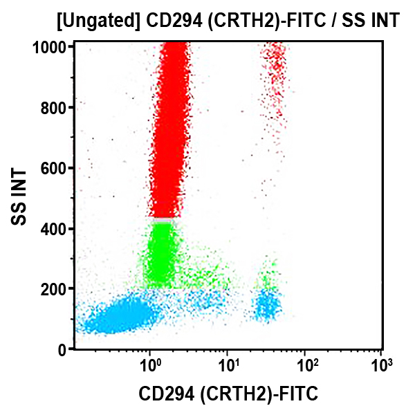
CD294 (CRTH2) Antibodies
CD294, also known as CRTH2, is a seven-transmembrane G-protein-coupled receptor known as the chemoattractant receptor-homologous molecule expressed on Th2 cells. Prostaglandin D receptor (DP), and CRTH2 have been identified as receptors for Prostaglandin D2 (PGD2), but they differ in their signaling pathways. PGD2 is the major metabolite of arachidonic acid produced by allergen-activated mast cells. CRTH2 molecule is preferentially expressed on human T-helper (Th) 2 and T cytotoxic (Tc) 2 cells but not on Th1 and Tc1 cells. CRTH2 is the most reliable surface marker selectively associated with circulating T (Th and Tc) cells able to produce IL-4 (as well as IL-5 and IL-13) and not IFNγ. On normal whole blood leucocytes, CD294 is also highly expressed on basophils and eosinophils. The expression of CD294 in peripheral blood mononuclear cells (PBMCs) is restricted to an activated state of Type 2 cells. CD294 may be weakly expressed on some monocytes and/or dendritic cells (CD14dim, CD16pos,HLA-DRpos,CD33pos). CD294 may act in mediating the recruitment and/or activation of basophils, eosinophils and Th2 cells at sites containing mast cells activated by invading allergens. CRTH2, but not DP, induces migration of Th2 cells, eosinophils and basophils in response to PGD2.
| Clone: BM16 | Isotype: IgG2a Rat |
| The BM16 monoclonal antibody precipitates a 55 to 70 kDa protein from cell lysates of CRTH2-transfected Jurkatt and Th2 clone, corresponding to PGD2 receptor. | |