Ki-67 Antibodies
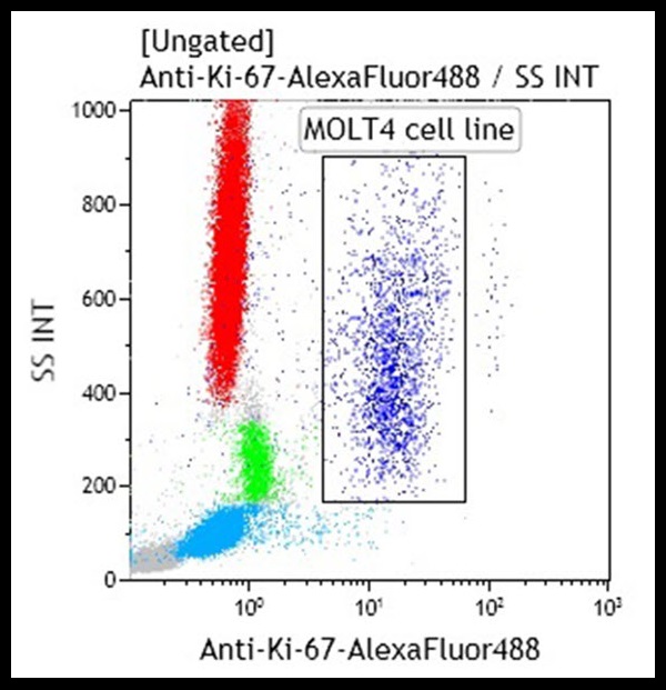
Ki-67 nuclear antigen is associated with cell proliferation and is found throughout the cell cycle (G1, S, G2, M phases). It is never expressed by resting (G0) cells. The Ki-67 antigen is a human nuclear protein defined by its reactivity with monoclonal antibody from the Ki-67 clone. Two isoforms of 345 and 395 kDa have been identified by cDNA sequence coding. Deduced amino acid sequence analysis of the Ki-67 antigen confirmed that the cDNA encodes for a nuclear and short-lived protein without any significant homology to known sequences. The Ki-67 antigen is expressed at active phases of the cell cycle (G1, S, G2 and M phases), but it is absent in resting cells (G0 phase). The level of Ki-67 antigen expression varies during cell cycle and it has been correlated to several cell pathways: The Ki-67 decrease pathway is characterized by a declining Ki-67 staining and leads eventually to the exit from the active cell cycle (G0). If cells on this pathway get stimulated by growth factors, they can enter the Ki-67 increase pathway that brings the cells back into S phase. Cells following the Ki-67 stable pathway exhibit a constant intensity of Ki-67 staining during the G1 phase. This pathway is thought to correspond to optimal local growth conditions. The cellular localization of the Ki-67 protein is cell cycle phase dependent. During interphase, the antigen can be exclusively detected within the nucleus, whereas in M phase most of the protein is relocated to the surface of the chromosomes. The antigen is rapidly degraded as the cell enters the non proliferative state.
| Clone: Ki-67 | Isotype: IgG1 Mouse |